Aim: Torpedo maculopathy (TM) is an incidental, congenital retinal lesion. The typical clinical finding is a unilateral, symmetric, oval, hypopigmented lesion in the inferotemporal macula. In most cases, the lesion is along the horizontal raphe, is torpedo-shaped, and the nasal edge is directed into the foveola. The diagnosis is determined on the basis of its characteristic shape, localization and findings on optical coherence tomography (OCT). The etiology and pathogenesis of torpedo maculopathy is unclear, but it is believed to be a congenital defect of the retinal pigment epithelium (RPE). The aim of this publication is highlight this diagnosis and to present an incidental finding of torpedo maculopathy in an adult patient.
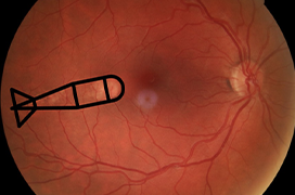
Case report: A 30-year-old female patient reported for a routine eye examination. Fundus examination of the right eye revealed an oval hypopigmented lesion with a size of 1 disk diameter inferotemporally from the fovea, which was followed by a satellite lesion in the same axis directed into the foveola. Based on OCT, OCT angiography, fundus autofluorescence, and the typical shape and location of the lesion, the patient was diagnosed with torpedo maculopathy in the right eye.
Conclusion: In general, torpedo maculopathy is an asymptomatic, congenital, benign retinal lesion, which is mostly diagnosed accidentally during a routine fundus examination. TM is non-progressive retinal finding with a minimal risk of deterioration of visual functions, which does not require any treatment. Nevertheless, due to the rare risk of a choroidal neovascular membrane, it is recommended to examine patients once a year. It is necessary to consider this diagnosis when a unilateral hypopigmented lesion is found inferotemporally from the fovea, and to distinguish it from chorioretinal atrophy, scar, vitelliform dystrophy, or other RPE lesions as part of the differential diagnosis.