Aim: We present a case report of a patient with dry form of age-related macular degeneration (AMD) in whom we monitored the effect of rheohemapheresis (RhF) treatment over 6 years.
Methods: A 67-year-old man was examined in April 2014 at the Department of Ophthalmology at the University Hospital in Hradec Králové for metamorphopsia and decreased visual acuity of the left eye. The patient received general treatment for hypercholesterolemia with Lipfix 10 mg once a day (atorvastatin 10 mg). The cholesterol level in the blood was 3.41 mmol/l, the lipid profile was normal. The patient’s previous ocular medical history was unremarkable.
The patient reported ocular complaints over the course of the last year, the main symptom of which was image distortion on the Amsler grid on the left eye. Baseline best corrected visual acuity of the left eye was 6/10. Visual acuity in the right eye was 6/6. In both eyes, the findings on the anterior segment corresponded to the patient’s age, with the exception of incipient cortical cataract. On the fundus of both eyes, the border of the optic nerve was demarcated, in the macula of the left eye there were a number of soft confluent drusen, in the central periphery there were no pathological changes. On the fundus of the right eye the finding was similar, but to a lesser degree. Optical coherence tomography on the macula of the left eye showed drusoid ablation of the retinal pigment epithelium (RPE), with accumulation of hyperreflectivities below the RPE. Pattern-reversal electroretinography (pERG): showed a slightly prolonged retention of the activity of the central region of the retina (p50 wave) and ganglion cells (N95 wave). Multifocal electroretinography (mfERG) in the central 30° of the retina was within normal limits. Electroretinography (ERG) demonstrated physiological photopic and scotopic rod activity. The patient was treated with 8 RhF procedures, two per week, with a 2-week interval, and the pulse was repeated 4 times.
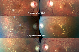
Results: We noted a gradual resorption of soft drusen in the patient, with attachment to the RPE line according to OCT examination at the following six-monthly check-ups over the next 6 years. Visual acuity of both eyes was maintained at the baseline values at the last check-up in April 2020, the area of soft drusen was significantly reduced. The function of the rod, cone system and the central region of the retina mfERG fluctuated only to an insignificant degree during the entire follow-up period, with a tendency towards a slight increase in activity after RhF treatment.
Conclusion: We noted an improvement of the anatomical and functional findings in a patient with dry form of AMD during a 6-year follow-up period after RhF treatment. The visual acuity of the affected eye remained at the baseline values.