
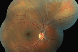


The review points to the issues of persistent hyaloid artery, more precisely to possible clinical features, the influence on visual functions and potential complications during intraocular surgeries. In professional journals we can find just few reviews regarding this rare deviation of the eye development, therefore we want to present our experience. The persistent hyaloid artery causes chronical local changes of eye background at both of our patients, retinal detachment and retinoschisis. The findings weren’t accompanied by significant decrease of visual functions or subjective patient’s complaints. Considering the potential complications published in journals such as hemoftalmus or retinal vessel occlusion we decided to be more conservative. That’s why we just checked-up the condition of the eye background and we were prepared to perform a surgery if necessary.
- OCT findings and long-term follow-up results of vitrectomy in patients with optic disc pit and associated maculopathy
- Persistent hyaloid artery - Indication for surgery or not?
- Combination of intravitreal corticosteroid with anti-VEGF in macular edema secondary to retinal vein occlusion
- Sarcoidosis and its ocular manifestations (an analysis of six case reports)
- Results of treatment of diabetic retionopathy by the Pascal laser system
- Ciliary body melanoma treatment by stereotactic radiosurgery
- Treatment results in patients with lymphoma disease in the orbital region
- The difference between the “Ganglion cell complex” and the retinal nerve fibre layer in the same altitudinal half of the retina in high-tension and normal-tension glaucomas
- Xerosis in patient with vitamin a deficiency
- Recommended procedure for eye examination for infants and children of pre-school age in regular outpatient practice