Introduction: Diagnostic and therapeutic management of the patient with malignant uveal melanoma (MMU) is subject to ongoing efforts to innovate. PET/CT (Positron Emission Tomography / Computed Tomography) examination is important in both diagnosis and metastases.
Material and methods: Evaluation of the importance of PET/CT examination in the group of patients diagnosed with MMU in the period 12.1.2016 to 6.12.2018. All patients with a diagnosis of secondary retinal detachment, suspected uveal melanoma, underwent standard examinations to detect possible metastases (liver ultrasound, chest X-ray). Patients for whom a stereotactic radiosurgery solution was planned due to the stage of the disease this examination was to exclude metastasis in the liver or lungs. PET/CT examination is part of the protocol within the exclusion criteria for treatment with stereotactic radiosurgery in one day session surgery.
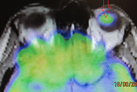
Results: In the group of 84 patients, 47 women (56 %) and 37 men (44 %) were aged between 26 and 90 years. Their average age was 61.4 years. The median group was 64 years, modus 65 years. Of 84 patients, 79 (94 % of cases) had a diagnosis of C69.3 (choroidal melanoma) and 5 patients (6 % of cases) had a diagnosis of C69.4 (ciliary body melanoma). Subsequent PET/CT examination in many patients did not reveal hypermetabolic manifestations that could involve various pathological processes, in others the radiopharmaceutical was captured in the primary tumor area of the uveal tract. Hypermetabolism in eye globe was only found in melanomas with a volume of more than 0.5 cm3. PET/CT examinations were 85, with one patient undergoing examination twice. However, in 25 patients (26 examinations), the radiopharmaceutical was taken up in places that subsequently required closer attention. The initial aim of the examination was to locate possible metastases of MMU. In the others, 3 incidents have been reported: increased metabolism in the lung and liver, thyroid and mediastinal lymph nodes. Of the 85 examinations, 26 (30.6 %) resulted in a hypermetabolic manifestation of accumulation, which was not located in the eye tract, resp. right in the eye. Two malignancies (prostatic carcinoma and rectosigmal carcinoma) have occurred in two patients. Very important was the discovery of MMU metastasis in the liver, which confirmed the important role of PET/CT examination in the management of MMU patients. The metastasis was discovered after repeated PET/CT examination.
Conclusion: PET/CT examination is a technically demanding examination and is one of the possibilities of imaging intraocular melanoma in tumors with volume more than 0.5 cm3. It is important in determining the grading and staging of the disease before radiosurgical treatment and also in detecting possible metastases after MMU treatment in cases where ultrasound or MRI examinations do not give a definite result. However, our study confirmed the significance of this examination for randomly detected 2 duplex malignancies (2.4%) and 3 incidentalomas (3.6%) in patients whose ophthalmologist diagnosed uveal melanoma and sent patients for full-body PET/CT examination.