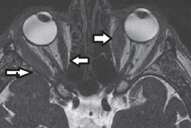
Due to the increased availability of MRI, this modality is the first choice for patients with a suspected pathology of the optic nerve, chiasm and optic tracts. Magnetic resonance imaging allows to evaluate the optic nerve itself as well as the gain or atrophy, its focal changes; it also allows detailed views of the surrounding structures such as vagina of the optic nerve and the mutual ratio between the full thickness of the nerve and the vagina, and the nerve itself. MR method uses a tissue contrast of an adipose tissue structures to a detailed imaging of the orbit. These data can play an important role not only in the diagnosis of the diseases with ophthalmic symptoms, but also in the diagnosis of the diseases of the nervous system. We are presenting a comprehensive overview of basic sequences used to show the optic nerve and the structures of the orbit as well as highlighting the benefits of their use and emphasizing their limitations. Imaging of the optic nerve and eye sockets may be standardized, and thus make the assessment easier for the following examinations that should be ideally performed using the same equipment and the same protocol display. The issue of imaging on the display unit with the strength of 1.5 Tesla is discussed; it is a machine that is largely represented across the Czech Republic.

