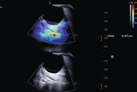
Shear wave elastography (SWE) is a new non-invasive diagnostic imaging technique, that maps the elastic properties of tissues. Nowadays this modality develops increasingly in medicine across its disciplines and opens a new era of high-quality ultrasound examination because it increases the specificity and thus improves diagnostic assurance. This method is similar to manual palpation, shows elastic properties of biological tissues and provides a kind of reconstruction of the internal structure of soft tissues based on measurement of the response of tissue compression. Various biological tissues have different elasticity and changes of these elastic properties often reflect pathological processes in the tissue and its abnormalities. This method is already used routinely on some foreign institutions in the detection and diagnosis of breast cancer and thyroid cancer, prostate cancer, in hepatology, cardiology, view the carotid arteries and lymphatic nodules. Finally examines its unquestioned benefit in ophthalmology. The output of elastography is an ultrasound image B-mode superimposed color-coded map. Shear waves elastography provides three major innovations: the quantitative aspect, the spatial resolution and the ability to run in real time.
- New Diagnostic Imaging Technique – Shear Wave Elastography
- Historic Survey of Posterior Lamellar Keratoplasty Techniques – Overview
- Ocular Surface Evaluation in Patients Treated With Prostaglandin Analogues Considering Preservative Agent
- Gene Therapy for Inherited Retinal and Optic Nerve Disorders: Current Knowledge
- Haemangiomas are Common Benign Tumors of the Child
- The Braf Mutation and the Possibilities of Uveal Melanoma Metastasing Prognostic Markers’ Identification
- Ocular Motility Disorders With Diplopia Like the First Symptoms Of Paranasal Tumours With Orbital Invasion – A Case Report