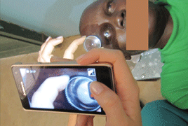
Documentation of the anterior segment and the eye fundus with instruments that enable quality precision diagnostics is a common and important part of screening in humanitarian ophthalmology projects. It is the essential element in diagnosis, monitoring and management of eye diseases. In sub saharan countries within the screening for ophthalmologist are not available the modern technologies such as biomicroscope (slit lamp) or fundus camera. We describe our experience with photographs of anterior segment of the eye by using digital camera and Smartphone. The documentation of the eye fundus was recorded through 20D Volk spherical lens to Smartphone.
Material and methods: Within the screening projects in collaboration with St. Elisabeth University of Health and Social Sciences for eye diseases in the year 2014 in Bigugu, Rwanda and in 2015 in Mapuordit, South Sudan, we examined patients who were unable to reach ophthalmologic care. We used a flashlight, a direct ophthalmoscope, tables to determine visual acuity on illiterate, Schiøtz tonometer, Volk lens, Smartphone. Patients who underwent screening, and needed glasses, got from humanitarian collection already used dioptric eyeglasses or sunglasses. For documentation of the anterior segment we used a digital camera and for patients in whom it was necessary to document fundus findings detected by direct ophthalmoscopy we took the opportunity of Smartphone with 8 Mpix camera and the LED
flash and Volk lens plus 20 Diopters.
Results: In 2014 within the project in Bigugu, Rwanda and in 2015 in Mapuordit, South Sudan, we examined patients in an improvised clinic without access to electricity. We examined in 2014 a total of 340 patients and in 2015 a total of 290 patients. Patient age was due to the unavailability of designated identification records estimated with the help of an interpreter. In both groups, the mean age of the patients was about 30 years. The most common diseases leading to blindness were cataract, trachoma, post-traumatic conditions. Infectious diseases and consequences of untreated infectious diseases were the cause of 20% of the permanent changes on the surface of the eye or the adnexa. In the group of HIV positive patients we did not mention pathological findings on the eye fundus.
Conclusion: Anterior segment findings documentation with digital camera or mobile phone and fundus examination using a Smartphone and Volks lens with a value of plus 20D is inexpensive and manageable technique which can capture high quality and reproducible images. These techniques are suitable for photo documentation of anterior segment and also eye fundus screening within humanitarian projects of eye diseases in developing countries.