
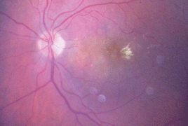
2015 - 2 Issue

Original article

Treatment of Pediatric Traumatic Macular Holes

Full Text links

Summary

Purpose: To report the results of treatment of the traumatic macular holes (TMH) in four children.

Methods: Retrospective study analyzed data of 4 children, males with a mean age of 12.3 years (range, 9–17 years), with diagnosis of TMH. All patients suffered a blunt trauma of the eye during the sport activities. The symptoms of three patients began after being hit to the face with a soccer balls, one boy was hit to his eye by a tennis rocket. Right eyes and left eyes were occurred identically. All patients were followed with ophthalmic examination, fundus photography and optical coherence tomography (OCT). One patient aged 10 years two weeks after blunt trauma with a soccer ball achieved spontaneous closure of TMH. Three patients aged nine to 17 years with TMH underwent surgical repair between September 2007 and May 2012 with three-port vitrectomy. After induction of posterior vitreous detachment vitrectomy with or without internal limiting membrane (ILM) peeling and gas or silicone oil injection were performed followed by prone positioning of head for ten days. Silicon oil was in an only patient removed within 3.5 month.

Results: All four macular holes were closed successfully. Follow-up period was from 10 to 31 month (ranged, 20 month). There were no reoperations. There were no surgical complications during follow-up period. Visual acuity (VA) improved in all eyes. In spontaneously closed TMH was VA improved from 0.5 to 1.0. In surgically treated group VA improved from initial 0.016 to 0.1 (ranged, 0.061) to final 0.25 to 0.5 (ranged, 0.36).

Conclusion: Pars plana vitrectomy is a safe method for treatment TMH in children without tends to spontaneous closure of TMD in OCT imaging. Predisposition for TMH in population south-east Asia is suspected.

References

  1. Bressler, S.B., Bressler, N.M., Fine, S.L. et al.: Natural course of choroidal neovascular membranes within the foveal avascular zone in senile macular degeneration. Am J Ophthalmol, 93(2); 1982: 157–163.
  2. Congdon, N., O’Colmain, B., Klaver, C.C. et al.: Causes and prevalence of visual impairment among adults in the United States. Arch Ophthalmol, 122 (4); 2014: 477–485.
  3. Heier, J.S., Brown, M.D., Chong, V. et al.: Intravitreal aflibercept (VEGF Trap- -Eye) in wet age-related macular degeneration. Ophthalmology, 119 (12); 2012: 2537–2548. of vascular endothelial growth factor (VEGF) and related ligands by VEGF Trap, ranibizumab and bevacizumab. Angiogenesis, 15(2); 2012: 171–185.
  4. Chang, A.A., Li, H., Broadhead, G.K. et al.: Intravitreal aflibercept for treatment-resistant neovascular age-related macular degeneration. Ophthalmology, 121; 2014: 188–192.
  5. Cho, H., Weber, M.L., Shah, C.P. et al.: Initial utilization of aflibercept in exudative age related macular degeneration. Eur J Ophthalmol, 24(4); 2014: 576–581.
  6. Martin, D.F., Maguire, M.G., Ying, G.S. et al: Ranibizumab and bevacizumab for neovascular age-related macular degeneration. N Engl J Med, 364(20); 2011: 1897–1908.
  7. Papadopoulos, N., Martin, J., Ruan, Q. et al.: Binding and neutralization
  8. Resnikoff, S., Pascolini, D., Etya’ale, D. et al.: Global data on visual impairment in the year 2002, Bull World Health Organ, 82(11); 2004: 844–851.
  9. Rudge, J.S., Holash, J., Hylton, D. et al.: VEGF Trap complex formation measures production rates of VEGF, providing a biomarker for predicting efficacious angiogenic blockade. Proc Natl Acad Sci U S A, 104(47); 2007: 18363–18370.
  10. Sawa, Y. et al.: Short-term outcomes of aflibercept for recurrent neovascular lar degeneration. Pharmacoeconomics, 24(4); 2006: 319–334.
  11. Schmidt-Erfurth, U. et al.: Intravitreal aflibercept injection for neovascular age-related macular degeneration. Ninety-six week results of the VIEW studies. Ophthalmology, 121; 2014: 193– 201.
  12. Schmier, J.K., Jones, M.L., Halpern, M.T.: The burden of age-related macu-
  13. Soubrane, G., Cruess, A., Lotery, A. et al.: Burden and health care resource utilization in neovascular age-related macular degeneration: findings of a multicountry study. Arch Ophthalmol, 125(9); 2007: 1249–1254.
  14. Eylea (aflibercept injekčný roztok v liekovke) Súhrn charakteristických vlastností lieku. Berlin, Nemecko: Bayer Pharma AG: 2014. age-related macular degeneration in Japanese patients. E-poster PO-1531, World Ophthalmology Congress 2014, Tokyo, Japan, April 2–6, 2014.
  15. Wykoff, C.C., Brown, D.M., Maldonado, M.E. et al.: Aflibercept treatment for patients with exudative age-related macular degeneration who were incomplete responders to multiple ranibizumab injections (TURF trial). Br J Ophthalmol, 98(7); 2014: 951–955.
  16. Yonekawa, Y., Andreoli, C., Miller, J.B. et al.: Conversion to aflibercept for chronic refractory or recurrent neovascular age- -related macular degeneration. Am J Ophthalmol, 156(1); 2013: 29–35. We hereby inform you that on the day of 26 March 2015, at a ceremony held in the Theresian hall of the Břevnov monastery, Dr. Jaroslava Vladyková was awarded the honour of Knighthood of Czech Medicine. After Professor Ivan Karel she is the second ophthalmologist to be awarded this prestigious honour. Journal editorial board NOTIFICATION