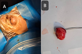
In this case report, we describe a 53-year-old woman who presented with a slow-growing lower lid mass in her right eye. On gross examination, a remarkable lower lid bulging was noted. On palpation, a subcutaneous oval-shaped mass with a firm consistency, measuring about 2cm, was noted. The uncorrected visual acuities of the patient were 20/20 (by Snellen chart) bilaterally, and the examinations of the anterior and posterior segments of both eyes were unremarkable. On the orbital Computed Tomography scan of the patient, a solitary and homogenous solid globular mass with the same density of the brain tissue was obvious. The patient underwent surgical excision. Microscopic assessment of the lesion revealed a biphasic hypercellular area (Antoni A) and myxoid hypocellular areas (Antoni B), containing slender cells with tapered ends, interspersed with collagen fibers, consistent with a diagnosis of schwannoma. In addition, some foci of nuclear palisading around the fibrillary process (Verocay bodies) could frequently be found throughout the highly cellular regions. Schwannomas rarely occur in the eyelids, but have clinical and paraclinical indicators which indicate the probable diagnosis. In conclusion, we suggest that eyelid schwannoma be considered as an element of the differential diagnoses list for subcutaneous lesions of the eyelid.
- Výpočetní tomografie a magnetická rezonance orbity v diagnostice a léčbě endokrinní orbitopatie – zkušenosti z praxe. Přehled
- Binokulární funkce před a po operaci strabismu u dospělých
- Vzdálená nasální část zorného pole – Část I
- Vzdálená nasální část zorného pole – Část II. Příspěvek k včasné diagnostice glaukomu
- Porovnání měření nitroočního tlaku u pacientů v inaktivní fázi endokrinní orbitopatie pomocí tří různých typů tonometrů
- Eyelid Schwannoma. A Case Report