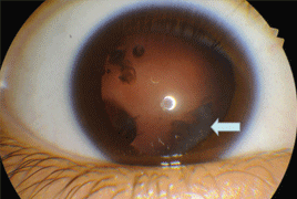
The authors present a case report of a three-year-old female patient with bilateral multiple anterior vitreous cysts. During examination for intermittent esotropia of the right eye was diagnosed not only hypermetropia, astigmatism and anisometropia, but also pigmented changes in peripheral retrolental space of both eyes. Clinical examination under general anaesthesia revealed bilateral multiple pigmented immobile vitreous cysts. There were five almost spherical, translucent, but slightly pigmented cysts on its cover on the right eye and four similar on the left, but visible only with dilated pupils. A dilating of pupils was slow and required more mydriatics than in similar aged children. Follow up period is ten years now. Occlusion therapy of amblyopia was performed to nine years of age. Treatment of refractive error and esotropia with correction for hypermetropia, astigmatism and anisometropia continues. Best corrected visual acuity in thirteen-year-old girl is 1,0 in both eyes without any visual disturbances described by patient. Corrected visual acuity in each eye is 1,0, right eye with +3,5 D sph., -3,5D cyl., axis 175°, left eye with +7,5 D sph., -3,0 D cyl., axis 35°. Patient is otherwise healthy and without any mental deficit. Position of all cysts remains unchanged and stabile during the follow up period (with recommendation to avoid hits to the head for all time, mainly in sports). Formation and slow progression of partial cortical cataract in the area of contact of the lens and one cyst in inferonasal quadrant of the lens on the right eye is monitored. A lamellar retinal extrafoveal defect of posterior pole of the right eye was found by OCT imaging.