Aims: To report a case of punctate inner choroidopathy (PIC) with pachychoroid disease features and active choroidal neovascular membrane.
Materials and methods: Case report
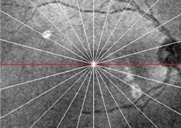
Results: A 33-year-old female patient with a history of myopic neovascular membrane in the right eye (OD), who had received multiple doses of intravitreal Aflibercept, consulted our retina service. Best-corrected visual acuity was 20/40 in OD. Fundus examination revealed small, well-defined, yellow-gray spots with subretinal fluid limited to the posterior pole. Optical coherence tomography scans from the OD showed subretinal fluid and a hyperreflective material between the epithelium/Bruch membrane (RPE/ BrM) complex, associated with loss of normal choroidal architecture and focal conformational choroidal excavation. There was also diffuse choroidal thickening in the macula, with pachyvessels compressing the inner choroid. Fundus autofluorescence showed active PIC lesions. With these findings, the diagnosis of punctate inner pachychoroidopathy associated with active choroidal neovascular membrane was made and Aflibercept therapy was restarted.
Conclusions: A subset of patients with PIC exhibits features associated with pachychoroid disease. This subtype of PIC is determined by unique demographics, multimodal image findings, and complications that differ from classic PIC, due to the potential influence of choroidal venous insufficiency on PIC manifestations and secondary complications.