Optical coherence tomography (OCT) has become a key tool in the differential diagnosis of optic neuropathies (ON), particularly in differentiating between glaucomatous and non-glaucomatous ON. Correct diagnosis is an essential factor for effective treatment management and prevention of progressive loss of vision. While glaucomatous ON is characterized by specific structural changes in the optic nerve head and retinal layers, non-glaucomatous neuropathies can be caused by a wide range of other causes, including inflammatory, ischemic or compressive processes. OCT allows visualization of the fine anatomical details of the optic nerve head and retina, providing valuable information for differential diagnosis. The importance lies in the physician’s ability to correctly interpret these images and integrate them into the patient’s overall clinical picture. This review focuses on the key features of glaucomatous and non-glaucomatous ON that can be detected early with OCT and highlights the importance of using this technique in everyday clinical practice.
- OCT in the Differential Diagnosis of Optic Neuropathies. A Review
- Cytomegalovirus Anterior Uveitis
- Clinical History Method versus Corneal Tomographers in Estimating Corneal Power after Photorefractive Surgery
- The Effect of Retinal Tear Location and Internal Tamponade on The Success of Pars Plana Vitrectomy in Patients with Uncomplicated Retinal Detachment
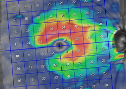
- The Relationship Between Retinal Nerve Fiber Thickness and Retinal Functional Sensitivity During Oct and Static Perimeter Examinations
- Internal Limiting Membrane Dehiscence and Rouleaux Formation in a Case with Branch Retinal Vein Occlusion and Macular Edema Treated with a Single Dexamethasone Implant Administration. A Case Report