Purpose: The problem of optic disc drusen (ODD) has been described in detail in several publications. However, less attention has been devoted to real haemodynamic parameters (HP) in ODD. It has been clinically demonstrated that the occurrence and progression of changes in the visual field in ODD are closely linked with the haemodynamics of the vascular supply of the eye – the optic nerve. ODD may visually overlap excavation of the disc of the optic nerve, on the basis of which it is more difficult to evaluate changes (scotomas) in the visual field in the case of glaucoma.
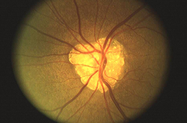
Methods: Haemodynamic parameters were prospectively evaluated in 54 patients with compensated intraocular pressure and with optic disc drusens. Drusens in the head of the optic nerve were demonstrated by a fundus examination and B-scan ultrasonography (USG). The drusens were divided into 3 groups according to the size of the individual drusens or drusen complex. Group I: area size up to 1.9 mm. Group II: area size: 1.9-3.9 mm. Group III: area size > 4.0 mm. Flow (haemodynamic) parameters – maximum systolic velocity (MSV), minimum diastolic velocity (MDV), and resistivity index (RI) and pulsatility index (PI) were recorded in the central retinal artery (CRA), in the central retinal vein (CRV), in the temporal and nasal ciliares posteriores arteries breves (CPAb) and in the ophthalmic artery (OA). The values were divided into 1. Physiological: CRA: 8.7 ± 0.9 / 2.9 ± 0.6 cm/s, or RI: 0.70 ± 0.05, 2. Slightly impaired: CRA: 6.6 ± 0.8 / 2.0 ± 0.5 cm/s, or RI: 0.75 ± 0.04. 3. Significantly impaired: CRA: 5.2 ± 1.2 / 1.9 ± 0.7 cm/s, or RI: 0.79 ± 0.03.
Results: No linear relationship was demonstrated between the size of the drusens and flow parameters. Slight impairment of HP in the CRA was present in 28.6% of drusens in group I, 48.3% in group II and 62.4% in group III. Significant impairment of HP in the CRA was present in 28.6% of drusens in group I, 48.3% in group II and 62.4% in group III. HP in the CPAb and OA were not of significant importance with regard to the presence and size of the drusens. The relationship between the individual variables was evaluated with the aid of a Pearson correlation coefficient: 0.213, group I P: 0.354, group II P: 0.073, group III P: 0.287.
Conclusions: HP are more often impaired in “large” optic disc drusens (group III), rarely in group I ODDs – though this is not an absolute rule. It is not possible to predict haemodynamic parameters according to the size of the drusen formation in the optic nerve. It appears that impairment of the haemodynamic parameters is conditioned not only by the size of the ODD, but also by the locality (distance from lamina cribriformis) and also the intrapapillary relationship to the vascular system.