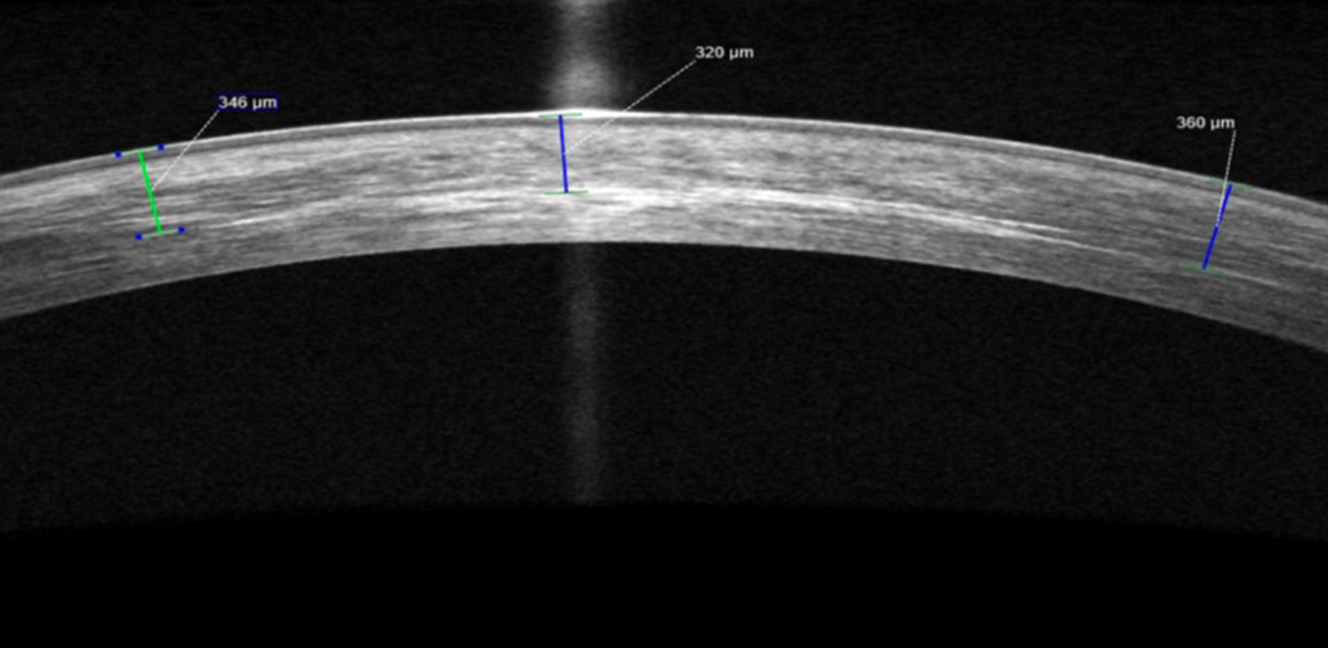
Objectives: Evaluation of the visibility and depth of the demarcation line in the corneal stroma in eyes with keratoconus 1 month and 3 months after epi-off accelerated corneal cross-linking (ACXL) using Anterior Segment Optical Coherence Tomography (AS OCT).
Material and Methods: This study analyses a group of 34 eyes with keratoconus 1 month and 3 months after ACXL (9 mW/cm2 for 10 min). The group was classified based on the ABCD clinical classification of keratoconus according to Belin and Duncan. AS OCT (Zeiss Cirrus 500, Anterior Segment Premier module) was used to assess the visibility and exact depth of the demarcation line in the corneal stroma.
Results: The demarcation line was visible 1 month after ACXL in 76.5 % of eyes with a mean depth of 238.13 ±20.36 μm and 3 months after ACXL in 100 % of eyes with a mean depth of 263.43 ±12.59 μm. Statistical analysis of the group did not show a significant relationship between the disease stage and the demarcation line visibility; however, there was a trend towards higher age (>30 years) in the group in those eyes where the demarcation line was visible vs. partially visible 3 months after ACXL. We found no difference in the mean and maximum line depth when comparing 1 month and 3 months after the procedure. There were no cases of disease progression 3 months after ACXL in the group.
Conclusion: Our study suggests that the assessment of the demarcation line in the corneal stroma is more reliable 3 months compared to 1 month after ACXL. We also observed a trend towards higher patient age in eyes where the demarcation line was clearly visible 3 months after ACXL. We did not confirm a relationship between the stage of keratoconus and the depth of the line, nor a difference in its mean and maximum depth 1 month and 3 months after the procedure.

